Delve into the world of cat scans, a cutting-edge medical imaging technique that has revolutionized healthcare. From its inception to its myriad applications, this comprehensive guide unveils the intricate details of cat scan meaning, providing a deeper understanding of this invaluable diagnostic tool.
As we delve into the technical aspects, we’ll explore the components of a cat scan machine, unravel the scanning process, and delve into factors that influence image quality. We’ll also shed light on the role of contrast agents, their types, uses, and safety considerations.
Overview of Cat Scan
A computed tomography (CT) scan, also known as a cat scan, is a medical imaging technique that uses X-rays and computer processing to create detailed cross-sectional images of the body. It provides more detailed information than traditional X-rays and is used to diagnose and monitor a wide range of medical conditions.
The basic principle of a cat scan is to rotate an X-ray beam around the body and measure the amount of radiation that passes through different tissues. This data is then processed by a computer to create cross-sectional images that show the internal structures of the body.
History of Cat Scan Development
The development of the cat scan began in the 1960s with the work of Godfrey Hounsfield, a British engineer. Hounsfield developed a prototype scanner that could produce cross-sectional images of the head. In 1979, Hounsfield and Allan Cormack, an American physicist, were awarded the Nobel Prize in Physiology or Medicine for their work on the development of the cat scan.
Medical Applications of Cat Scans
Cat scans have a wide range of medical applications, including:
- Diagnosing and monitoring cancer
- Evaluating injuries and trauma
- Detecting and monitoring heart disease
- Diagnosing and treating strokes
- Evaluating kidney and liver function
- Planning for surgery and other medical procedures
Technical Aspects of Cat Scan
The technical aspects of a cat scan involve the components of the machine, the scanning process, and factors affecting image quality.
The machine comprises an X-ray source, a rotating gantry, and an array of detectors. The X-ray source emits a narrow beam of X-rays that passes through the patient and is detected by the detectors on the opposite side of the gantry.
Scanning Process
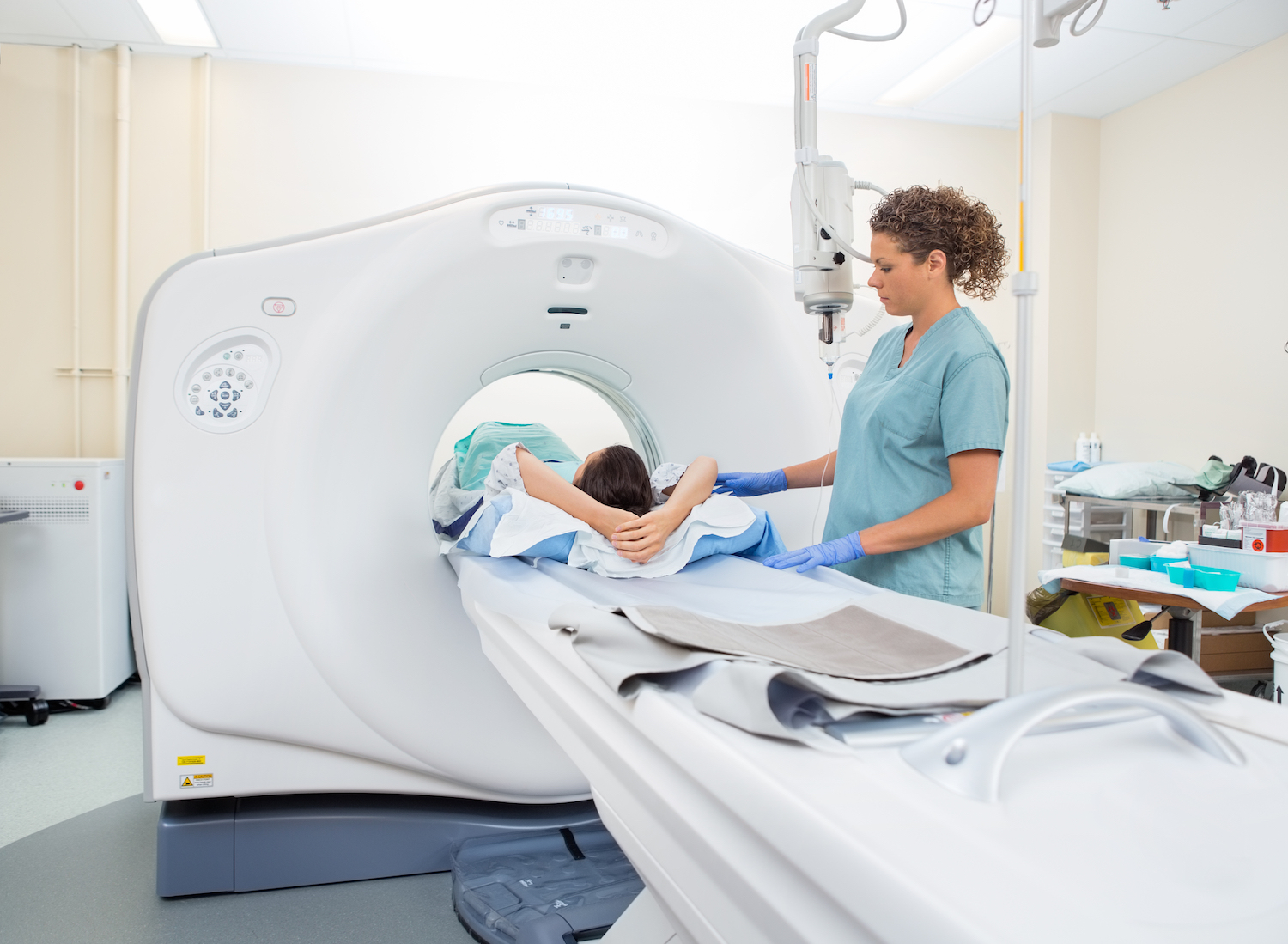
The scanning process begins with the patient lying on a table that moves through the gantry. The X-ray source and detectors rotate around the patient, collecting data from multiple angles. The data is then processed by a computer to create cross-sectional images of the body.
Image Quality
Image quality in a cat scan is influenced by factors such as the X-ray beam intensity, the number of detectors, and the reconstruction algorithm used. Optimization techniques like iterative reconstruction and noise reduction can improve image quality and reduce artifacts.
Contrast Agents for Cat Scans

Contrast agents are substances that are injected into the body before a cat scan to help improve the visibility of certain tissues and organs. They work by absorbing X-rays, which makes them appear brighter on the scan images.
There are two main types of contrast agents used in cat scans: iodinated contrast agents and gadolinium-based contrast agents.
Iodinated Contrast Agents
Iodinated contrast agents are the most common type of contrast agent used in cat scans. They are made up of iodine, a heavy element that absorbs X-rays well. Iodinated contrast agents are typically injected into a vein in the arm.
Iodinated contrast agents are generally safe, but they can cause some side effects, such as nausea, vomiting, and hives. In rare cases, they can cause a more serious allergic reaction called anaphylaxis.
Gadolinium-Based Contrast Agents
Gadolinium-based contrast agents are another type of contrast agent that is used in cat scans. They are made up of gadolinium, a rare earth metal that absorbs X-rays even better than iodine. Gadolinium-based contrast agents are typically injected into a vein in the arm.
Gadolinium-based contrast agents are generally safe, but they can cause some side effects, such as nausea, vomiting, and headache. In rare cases, they can cause a more serious condition called nephrogenic systemic fibrosis (NSF), which can lead to kidney failure.
Interpretation of Cat Scan Images

Interpreting cat scan images involves a systematic analysis of the grayscale images produced by the scanner. Radiologists, who are medical doctors specializing in interpreting medical images, play a crucial role in this process.
Common Anatomical Structures and Their Appearance on Cat Scans
Common anatomical structures visible on cat scans include:
- Bones:Appear as dense, white structures.
- Soft tissues:Appear as shades of gray, with different densities indicating different types of tissue (e.g., muscle, fat, organs).
- Air-filled structures:Appear as black or very dark gray areas (e.g., lungs, sinuses).
- Fluid-filled structures:Appear as dark gray areas (e.g., blood vessels, cysts).
Role of Radiologists in Interpreting Cat Scan Images
Radiologists use their expertise and knowledge of anatomy and pathology to:
- Identify and locate anatomical structures.
- Detect abnormalities in size, shape, or density of structures.
- Differentiate between normal and abnormal findings.
- Correlate findings with patient history and symptoms.
- Provide a written report and recommendations for further evaluation or treatment.
Applications of Cat Scans
Cat scans, also known as computed tomography (CT) scans, are a valuable diagnostic tool in modern medicine. They provide detailed cross-sectional images of the body, enabling healthcare professionals to diagnose and monitor a wide range of medical conditions.
Specific Medical Conditions Diagnosed Using Cat Scans
- Cancer: Cat scans can detect and characterize tumors, determine their extent, and monitor their response to treatment.
- Cardiovascular disease: Cat scans can visualize the heart, arteries, and veins, aiding in the diagnosis of conditions such as coronary artery disease and aortic aneurysms.
- Neurological disorders: Cat scans can assess the brain, spinal cord, and other nervous system structures, helping diagnose conditions like stroke, dementia, and multiple sclerosis.
- Musculoskeletal disorders: Cat scans can evaluate bones, joints, and muscles, aiding in the diagnosis of fractures, dislocations, and arthritis.
- Infections: Cat scans can detect and localize infections in various parts of the body, including the lungs, abdomen, and pelvis.
Advantages and Limitations of Cat Scans Compared to Other Imaging Techniques, Cat scan meaning
Cat scans offer several advantages over other imaging techniques:
- High resolution: Cat scans provide detailed images that allow for accurate visualization of anatomical structures.
- Non-invasive: Cat scans do not involve invasive procedures, making them a relatively safe and comfortable examination.
- Versatile: Cat scans can be used to image a wide range of body parts and can be combined with contrast agents to enhance the visibility of certain structures.
However, cat scans also have some limitations:
- Radiation exposure: Cat scans involve exposure to ionizing radiation, which can be a concern, especially for repeated examinations.
- Motion artifacts: Movement during the scan can result in blurred or distorted images, affecting the accuracy of the results.
- Contrast agents: Some cat scans require the use of contrast agents, which can cause allergic reactions in certain individuals.
Use of Cat Scans for Surgical Planning and Treatment Monitoring
Cat scans play a crucial role in surgical planning and treatment monitoring. They provide:
- Preoperative planning: Cat scans can help surgeons visualize the surgical area, plan the approach, and anticipate potential challenges.
- Intraoperative guidance: Cat scans can be used during surgery to provide real-time images, assisting surgeons in navigating complex procedures.
- Treatment monitoring: Cat scans can be used to assess the response of tumors or other conditions to treatment, allowing healthcare professionals to adjust treatment plans as needed.
Safety Considerations for Cat Scans
Computed tomography (CT) scans, commonly known as cat scans, involve the use of X-rays to create detailed images of the body’s internal structures. While CT scans are valuable diagnostic tools, it is important to be aware of the associated radiation exposure and the potential risks and benefits.
Radiation Exposure
CT scans use ionizing radiation, which has the potential to damage cells and increase the risk of cancer. The amount of radiation exposure varies depending on the specific scan performed and the size of the body part being imaged. However, CT scans generally involve a higher radiation dose than traditional X-rays.
Risks and Benefits
The risks and benefits of CT scans must be carefully weighed in each individual patient. The benefits of CT scans include the ability to diagnose a wide range of medical conditions, including cancer, heart disease, and stroke. However, the potential risks of radiation exposure must also be considered.
The risk of developing cancer from a single CT scan is generally considered to be low, but it increases with repeated scans and higher radiation doses. Children and young adults are more susceptible to the effects of radiation than older adults.
Minimizing Radiation Exposure
There are several steps that can be taken to minimize radiation exposure during CT scans:
- Only undergo CT scans when medically necessary.
- Discuss the potential risks and benefits with your doctor before undergoing a CT scan.
- Choose a facility that uses the latest technology and techniques to minimize radiation exposure.
- Consider alternative imaging techniques that involve less radiation, such as ultrasound or MRI.
By following these guidelines, you can help minimize the potential risks associated with CT scans and ensure that the benefits outweigh the risks.
Future Developments in Cat Scan Technology
:max_bytes(150000):strip_icc()/189603_color1-5bc4e2a246e0fb002697e9c1.png)
Cat scan technology is constantly evolving, with new advancements emerging to enhance image quality, reduce radiation exposure, and improve diagnostic capabilities.One significant development is the introduction of multi-detector computed tomography (MDCT), which utilizes multiple detectors to capture more data in a single scan.
This results in faster scan times, improved spatial resolution, and reduced noise.
Artificial Intelligence in Cat Scan Interpretation
Artificial intelligence (AI) is playing an increasingly important role in cat scan interpretation. AI algorithms can analyze large volumes of medical images to identify patterns and abnormalities that may be missed by the human eye. This can assist radiologists in making more accurate and timely diagnoses.AI-powered
software can also automate image reconstruction and processing, reducing the time required for image analysis and interpretation. This can improve efficiency and reduce turnaround times for patient results.
Wrap-Up: Cat Scan Meaning
In conclusion, cat scans have become an indispensable tool in modern medicine, offering unparalleled insights into the human body. Their versatility extends across a wide range of medical conditions, enabling precise diagnoses, surgical planning, and treatment monitoring. As technology continues to advance, we can anticipate even more groundbreaking applications of cat scans, further enhancing their role in healthcare.
FAQs
What is the principle behind a cat scan?
A cat scan utilizes X-rays and advanced computer processing to generate detailed cross-sectional images of the body, providing a comprehensive view of internal structures.
How long does a cat scan typically take?
The duration of a cat scan varies depending on the area being scanned and the complexity of the procedure, but it generally ranges from a few minutes to half an hour.
Are cat scans safe?
Cat scans involve exposure to ionizing radiation, but the radiation doses used are carefully controlled to minimize risks. The benefits of cat scans generally outweigh the risks, especially when used judiciously and when alternative imaging methods are not suitable.